Title: How to Make an AFM Sample of Graphene Quantum Dots
(how to make afm sample of graphene quantum dots)
Introduction:
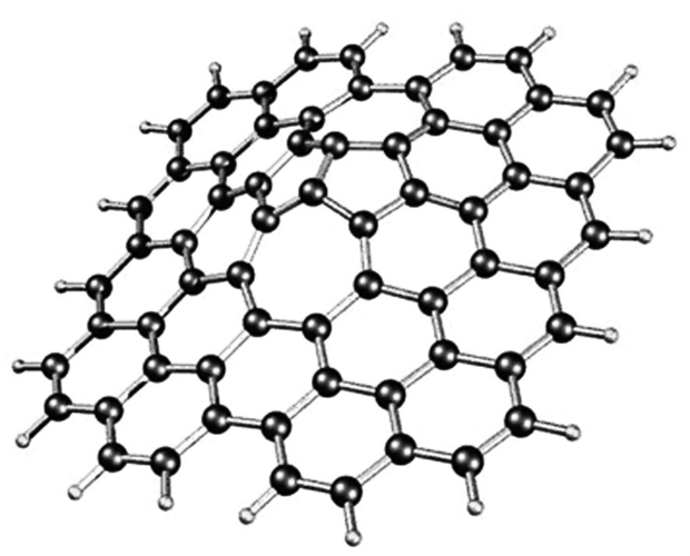
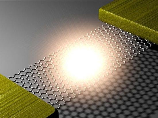
Graphene, a two-dimensional material with unique properties, has attracted significant attention in recent years due to its potential applications in various fields such as electronics, energy storage, and medicine. One promising application is the development of quantum dots, which can be controlled using light. In this blog, we will discuss how to create an AFM (Atomic Force Microscope) sample of graphene quantum dots.
Materials and Equipment:
Before starting the process, you will need to gather the following materials and equipment:
1. Graphene samples – You will need at least two sets of graphene samples for this project. The samples should be pre-grown on substrates such as silicon or metal.
2. AFM sample holder – This is an essential piece of equipment that holds the graphene sample during the AFM operation. It should be made from conductive material such as silver or gold.
3. AFM cantilevers – These are used to precisely align the graphene on the substrate and collect the image data.
4. Image sensor – A high-resolution image sensor such as a CMOS or complementary metal oxide semiconductor (CMOS) pixel camera is required for capturing the AFM image.
5. Image processing software – Software such as MATLAB or Python are needed for analyzing the AFM images and extracting information about the graphene quantum dots.
6. Light source – A laser with a wavelength of around 780 nm is commonly used to generate light for AFM experiments.
Procedure:
Step 1: Prepare the graphene samples – Place one graphene sample on the substrate and another on top of it. Ensure that both samples are clean and free of impurities. To achieve this, use a cleaning solution containing water and an acid such as HCl or HNO3. Rinse the graphene sample thoroughly before transferring it onto the substrate.
Step 2: Set up the AFM sample holder – Attach the AFM sample holder to the table or work station using strong electrical connections. Choose a sample holder with appropriate surface roughness for the AFM method.
Step 3: Align the graphene – Use the AFM cantilevers to align the graphene sample on the substrate. Perform multiple alignment cycles to achieve a stable configuration of the graphene on the substrate.
Step 4: Collect the AFM image – Use the high-resolution image sensor to capture the AFM image. The AFM image should be captured in grayscale mode to improve contrast and readability.
Step 5: Analyze the AFM image – Use image processing software to analyze the AFM image and extract information about the graphene quantum dots. Some of the key features of graphene quantum dots include their size, shape, and density distribution. Use the AFM image data to determine these features accurately.
Conclusion:
(how to make afm sample of graphene quantum dots)
In conclusion, creating an AFM sample of graphene quantum dots is relatively straightforward and requires only a few steps. By carefully selecting and preparing the graphene samples, setting up the AFM sample holder, aligning the graphene, collecting the AFM image, and analyzing the AFM image, you can gain valuable insights into the unique properties of graphene quantum dots. With the rapid advances in technology, there is tremendous potential for the development of new applications utilizing these quantum dots.


